Georgia Tech, Emory team creates open-source tool that lets researchers use artificial intelligence to analyze moving and still images collected by any imaging device
By Joshua Stewart
The last few decades have brought advances in biomedical imaging that allow researchers to capture still and moving images at an unprecedented level of detail. Analyzing those images, however, often remains a manual, error-prone process that fails to maximize their value for understanding biological systems.
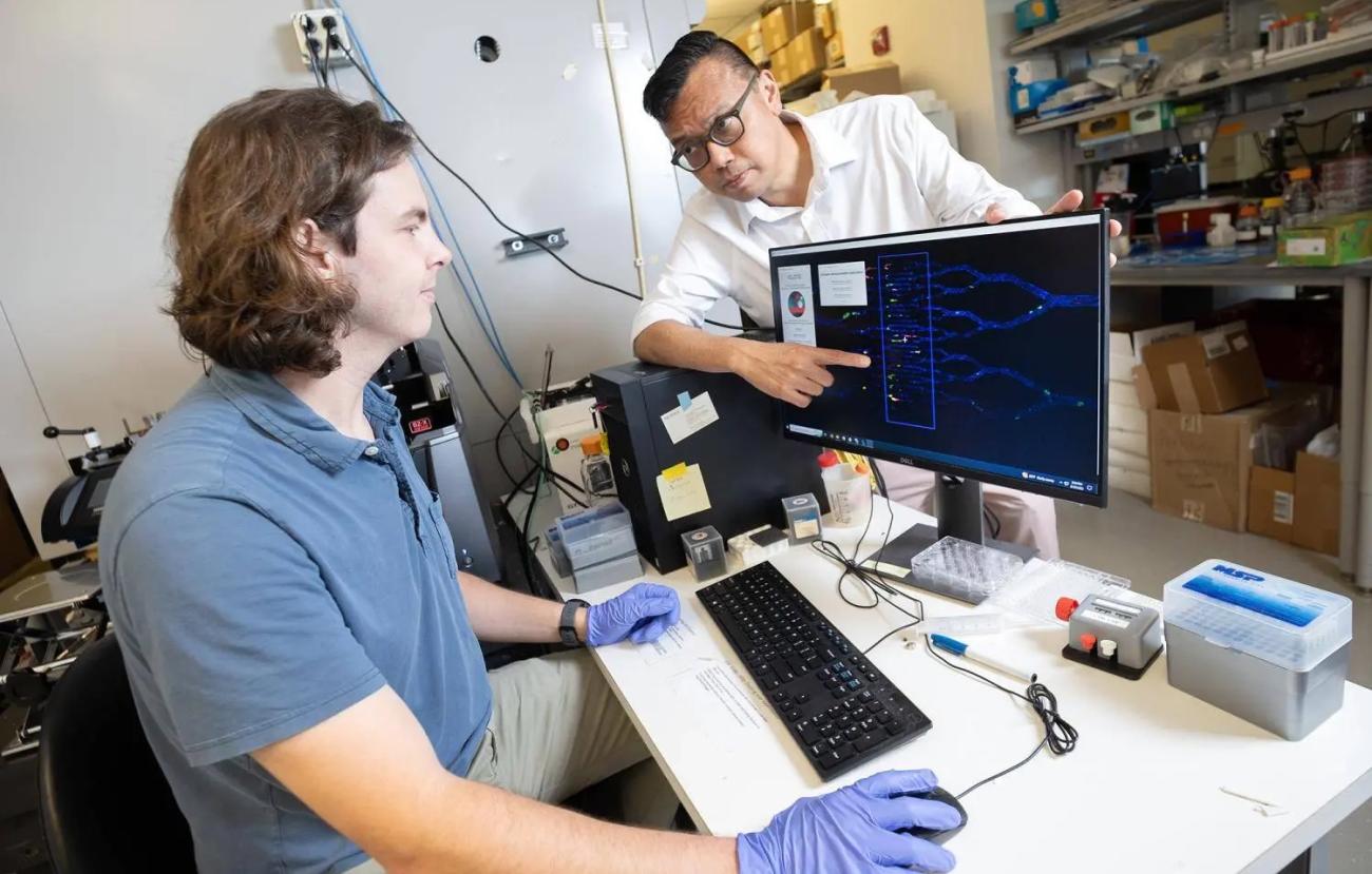
A team at Georgia Tech and Emory University has created a simple-to-use software program to help. It allows any researcher with imaging data to leverage powerful artificial intelligence algorithms and uncover new insights from their experiments — without knowing how to write complex computer code.
Called iCLOTS, the program is open-source and freely available on a dedicated website with extensive documentation and guidance. The team described the software and how it can help deepen understanding of experimental data in the journal Nature Communications.
“So many mathematicians and data science engineers are making really wonderful algorithms. And there are clinicians and researchers who could use them. But there’s a disconnect — the people who need them most really aren't able to access them,” said Meredith Fay, who led creation of iCLOTS and finished her Ph.D. in the Wallace H. Coulter Department of Biomedical Engineering at Georgia Tech and Emory in May.
“They know so much about hematology, medicine, or bioengineering, and it's not a great use of their time to learn to code and make software,” she said. “We’ve put all sorts of image processing and artificial intelligence algorithms into this simple interactive package so that anyone can use these methods.”
Fay is first author on the study with a team on both campuses and her Ph.D. advisor Wilbur Lam, who admitted existing software solutions are so complex, he can’t even use them.
“Meredith has found that nice balance where users are introduced to enough parameters they can adjust so the software can generalize to any type of image or video obtained from any type of microscopy equipment,” said Lam, a Coulter BME professor and associate dean for innovation in the Emory School of Medicine. “At the same time, it's not so burdensome that they have to read a manual or get a Ph.D. to learn how to use it.”
Other analysis software available to biomedical researchers typically works for one specific experimental or microscopy system. And even then, using the most powerful capabilities of the system requires significant expertise in software coding.
Lam said they made iCLOTS to complement and democratize those capabilities, not to compete with existing tools. They also made it open source in hopes others will build on the tool to make it even better for the biomedical research community.
The software adapts existing and well-validated algorithms, including some of the same methods used to guide self-driving cars. While designed to work with any imaging data, the researchers focused mostly on microfluidics platforms — typically small computer chip-like devices with micro- or nanoscale channels used to study cells or biofluids in motion. Complex dimensions and constantly moving samples make these experiments among the trickiest to study.
“Video analysis is a newer problem, and not many people have cracked it,” Lam said. “It’s not easy to do sophisticated analysis because those images are moving. Meredith was able to take these existing algorithms and apply them in a very usable fashion.”
To validate the tool’s effectiveness, Fay ran it through a battery of repeatability and sensitivity tests. The team also tried it on a variety of published datasets and compared the software’s analysis to the studies’ results. And they compared iCLOTS’ analyses to manual results compiled by humans looking at images or videos.
During the validation testing, the software uncovered insights in data from various projects in Lam’s lab that might otherwise have been missed. For example, the team reported new observations of mechanical properties of blood cells in sickle cell disease and changes in blood velocity in sepsis patients that indicated changes in blood viscosity.
“Some of this was enabled by the increased resolution that iCLOTS can provide: Not only did my coworkers know there was more of a particular clinical type of cell, they knew those cells were also smaller and had different morphology,” said Fay, who’s now a postdoctoral researcher at Duke University. “We make the argument in the paper that all this additional detail can enable a new level of hypotheses.”
Lam said that demonstrates the potential of their tool to advance research: “It goes back to how the standard for a lot of this is all manual right now. It's ironic, right? You have this high-tech experimental device, but the analysis is by hand. By virtue of using our software, you get so much interesting data and so many cool ways to interpret the data. The software itself could help us generate hypotheses that could advance and extend research.”
This research was supported by the National Institutes of Health Institute of Health Lung and Blood, grant Nos. R01HL130918, R01HL140589, R35HL145000, 5T32HL139443-03, HL160210-01, T32HL139443-3, and L40HL149069, and the National Institute of Allergy and Infectious Diseases, grant No. R38AI140299. Any opinions, findings, and conclusions or recommendations expressed in this material are those of the authors and do not necessarily reflect the views of any funding agency.
Citation: Fay, M.E., Oshinowo, O., Iffrig, E. et al. iCLOTS: open-source, artificial intelligence-enabled software for analyses of blood cells in microfluidic and microscopy-based assays. Nat Commun 14, 5022 (2023). https://doi.org/10.1038/s41467-023-40522-4
Latest BME News
Jo honored for his impact on science and mentorship
The department rises to the top in biomedical engineering programs for undergraduate education.
Commercialization program in Coulter BME announces project teams who will receive support to get their research to market.
Courses in the Wallace H. Coulter Department of Biomedical Engineering are being reformatted to incorporate AI and machine learning so students are prepared for a data-driven biotech sector.
Influenced by her mother's journey in engineering, Sriya Surapaneni hopes to inspire other young women in the field.
Coulter BME Professor Earns Tenure, Eyes Future of Innovation in Health and Medicine
The grant will fund the development of cutting-edge technology that could detect colorectal cancer through a simple breath test
The surgical support device landed Coulter BME its 4th consecutive win for the College of Engineering competition.

