Simons Foundation award supports research of information flow along the visual neural highway
By Jerry Grillo
In people with autism, neural “traffic jams” may slow the flow of information along visual routes in the brain, delaying timely, responsive actions.
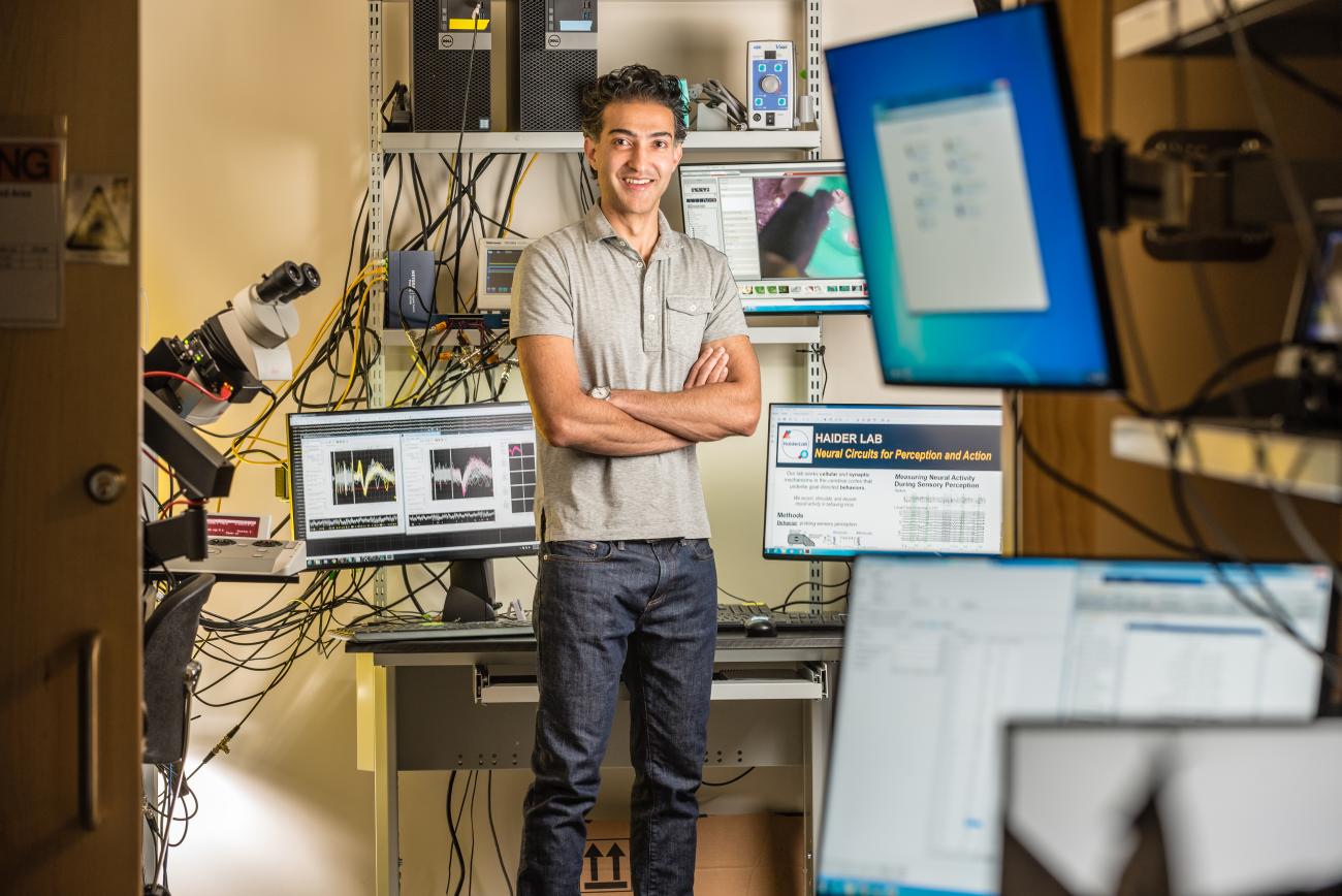
Just as highway departments study vehicle traffic flow, Georgia Tech and Emory researcher Bilal Haider is studying neural traffic in the brain to understand the holdups, and his discoveries could lead to therapeutic techniques to “unjam” the flow of neural information.
Now the Simons Foundation Autism Research Initiative (SFARI) is supporting his efforts. In December, Haider received one of 17 SFARI Pilot Awards, part of a $5 million national initiative supporting exploratory ideas with the potential to transform autism spectrum disorder research.
“This will be one of the first studies to test the idea that, because ‘traffic’ is slowed at multiple points in the brain, visual messages never get to the final destination to cause an action on time,” said Haider, assistant professor in the Wallace H. Coulter Department of Biomedical Engineering at Georgia Tech and Emory University. “Typical studies only focus on one area at a time. This one will be among the first to try and connect all the major dots along the route.”
Haider’s lab will use the three-year, $300,000 grant to comprehensively measure neural activity in mice while they view, process, and respond to visual stimuli. The idea, Haider said, is to develop a full picture of visual brain area activity using neural probe technology developed in part by his former colleagues at University College London. The probes will target each visual destination on the map to track electrical firing along the neural route.
The team will first identify a network of specialized visual brain areas — up to eight distinct regions — using a 20-year-old standard technique called blood flow imaging. The researchers have developed a way to make the skull transparent and image brain blood flow, basically using glass windows and a compound called cyanoacrylate.
“It’s Krazy Glue, essentially, and it keeps the skull healthy and intact so that we can later make really stable and uncompromised neural recordings with electrical probes in all of the destinations on the map, across several days or even weeks of recordings,” Haider said.
This kind of investigation with delicate neural probes in the brain would be difficult if the skull was removed in a mouse that is awake, breathing, and moving while performing a visual task.
The imaging tracks the amount of oxygen in blood by measuring how much light gets reflected back from the brain. So, with a high-speed camera and a bright red light focused on the brain surface, Haider’s team will measure the active versus inactive areas of the brain that demand oxygenated blood.
Using this system while showing the mouse strong flashing and moving visual stimuli, the researchers can find the active visual areas of the brain and build a map of the visual brain areas. The team will study both typical, wildtype mice, and also mice that have been engineered to have the same genetic mutation as some people with autism.
“Our goal is to measure how networks of neurons across these eight brain areas might miscommunicate, or get lost along the route, and lead to impairments of visual perception in the autism model mice,” Haider said. “We expect that the flow of visual information — the neural ‘traffic’ on the map — during visual task performance in the autism model mice will be slower and less orderly than in the typical wildtype mice.”
The findings of this research could help in the development of new therapeutic techniques to “unjam” the traffic at the key points of the route and speed up or restore behavioral performance.
“A longer-term application would be to connect with clinicians and human brain researchers who study individuals with autism,” Haider said. “Then we could see if EEG or functional MRI activity in people with autism also shows evidence of neural traffic jams along the same visual routes, and if these correlate with visual disturbances or poorer visual behaviors. We could also test promising therapeutics in mice to see if they ‘unjam’ traffic and improve behavior.”
Latest BME News
Jo honored for his impact on science and mentorship
The department rises to the top in biomedical engineering programs for undergraduate education.
Commercialization program in Coulter BME announces project teams who will receive support to get their research to market.
Courses in the Wallace H. Coulter Department of Biomedical Engineering are being reformatted to incorporate AI and machine learning so students are prepared for a data-driven biotech sector.
Influenced by her mother's journey in engineering, Sriya Surapaneni hopes to inspire other young women in the field.
Coulter BME Professor Earns Tenure, Eyes Future of Innovation in Health and Medicine
The grant will fund the development of cutting-edge technology that could detect colorectal cancer through a simple breath test
The surgical support device landed Coulter BME its 4th consecutive win for the College of Engineering competition.


