Capstone team publishes feasibility study on semiautomated device for scoliosis correction surgery
With placement of a few markers and a quick picture, a group of recent biomedical engineering graduates has created a way to quickly and accurately measure spinal alignment during surgery to correct scoliosis.
Their device uses computer vision and a kind of machine learning known as deep learning to calculate two critical measurements, and they’ve documented the feasibility of their approach in the Journal of Neurosurgery: Spine with mentors from Mayo Clinic in Jacksonville. Their study appeared online in early June.
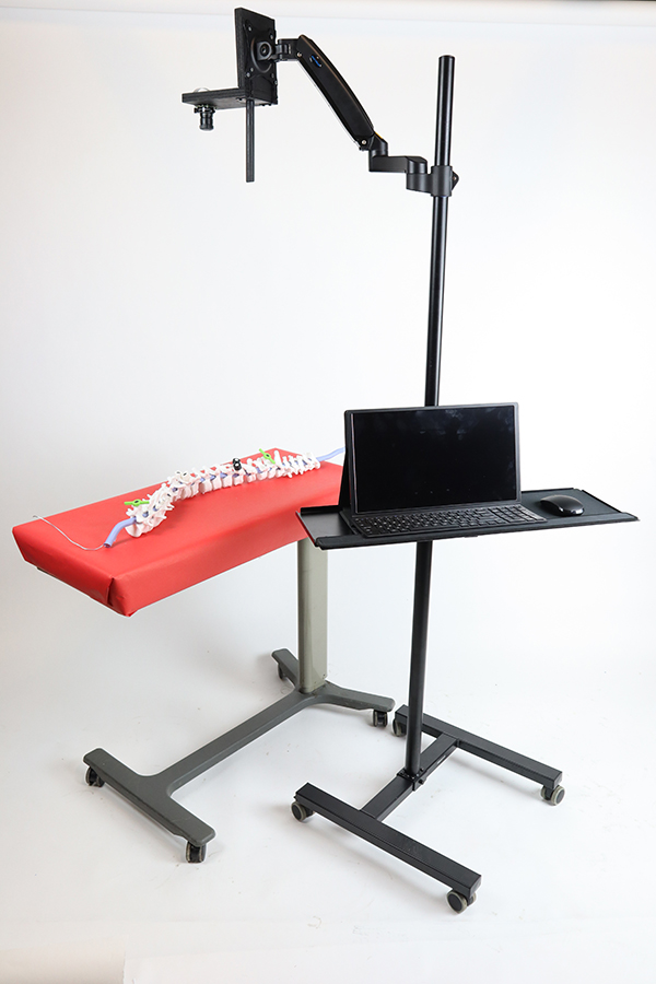
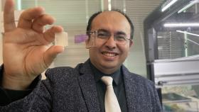
The prototype device built by a team of Capstone students in Spring 2021 to quickly and accurately measure spinal alignment during surgery to correct scoliosis without using radiographic approaches. The device uses an off-the-shelf camera, 3D-printed markers, and a custom deep learning algorithm to calculate two critical data points for doctors mid-surgery, the plumb line and Cobb angle. The team published a feasibility study on their work in the Journal of Neurosurgery: Spine. (Photo Courtesy: Yoel Alperin, Parth Gami, Sindhu Kannappan, and Kelly Qiu.)
Based on their work in Capstone Design in Spring 2021, the idea is to quickly and accurately give surgeons data on how well they have corrected the curvature of the patient’s spine without traditional radiographic tools. Usually that information would come via X-ray or CT scan in the operating room, with doctors pausing the procedure to acquire key metrics, like the Cobb angle and plumb line.
“There's currently not a convenient way to measure, intraoperatively, the alignment of the spine during scoliosis surgery,” said Kelly Qiu, who graduated in 2021 and was a member of the team. “The current methods are radiographic; however, because they are radiographic, they take a long time to take an image of the spine during the surgery, and it also exposes the patient as well as the staff in the OR to a lot of radiation.”
The group is the third Capstone team in the Wallace H. Coulter Department of Biomedical Engineering to publish their work in a peer-reviewed journal in the last year, a first for the program. Along with Qiu, the team included Yoel Alperin, Parth Gami, and Sindhu Kannappan, who all finished their bachelor’s degrees in 2021.
The measurement device relies on custom 3D-printed markers placed at key locations on the spine during surgery. An off-the-shelf camera captures an image, and a custom deep learning algorithm spots the markers and makes quick calculations for surgeons. Deep learning builds layers of algorithms in networks modeled on the human brain.
The team validated the system using a cadaver with scoliosis and reported the automated approach produced data well within clinically accepted parameters. The study also noted the prototype device could cut time for intraoperative measurement from 15 minutes for a typical radiographic approach to roughly one minute.
“We really took advantage of these brand new computational processing techniques. The fact that we're able to use a deep learning algorithm with image processing allows us to get rid of that need for massive radiographic instrumentation [in the operating room] like X-ray or CT machines,” Gami said, explaining that their relatively simple device is easy to operate and maneuver in the sterile operating room environment.
All of that streamlining could reduce costs for patients along with reducing risks of infection and exposure to radiation, Alperin said.
“There are huge savings on the actual device itself, and on top of that, anytime that you bring another person into the operating room, that's additional cost to the patient,” he said. “You have to bring in that person to actually run the [imaging] arm. And then there’s additional costs from the extension of the length of surgery.”
The project was personal for Alperin. He has a mild form of scoliosis, and while he’s never had to have surgery or wear corrective braces, he has long been part of a community of people who did. He knew the potential impact of the intervention, and he was invested in the effort to improve the process.
The group members said they hope work can continue on their approach now that they’ve demonstrated the feasibility and published their data — perhaps by medical device companies, other labs, or even a future Capstone team.
Gami noted improving the algorithm to identify the heads of the screws placed in the spine during surgery could eliminate the need for special markers, and there’s potential to integrate the system with cameras already present in the operating room. The team also noted they proved their system on spinal deformation in one plane and would need further work to validate its effectiveness for patients with two- or three-dimensional curvatures.
“The reason we wanted to publish this was that we know this works, and that deep learning approaches are underutilized in the ortho-spinal surgical area,” Gami said. “This could inspire people who have the resources and the time to advance our ideas even further.”
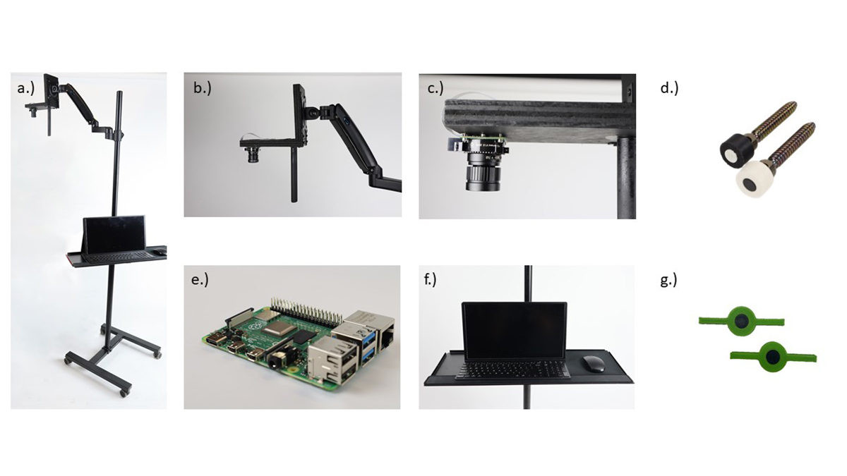
A figure from the journal paper breaking down the pieces of the team’s device to semi-automatically measure spine alignment during scoliosis correction surgery. The prototype uses a camera, 3D-printed markers placed along the spine (images marked D and G), and a custom deep learning algorithm. (Photo Courtesy: Yoel Alperin, Parth Gami, Sindhu Kannappan, and Kelly Qiu.)
Latest BME News

Jo honored for his impact on science and mentorship
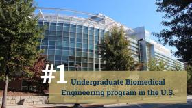
The department rises to the top in biomedical engineering programs for undergraduate education.
Commercialization program in Coulter BME announces project teams who will receive support to get their research to market.

Courses in the Wallace H. Coulter Department of Biomedical Engineering are being reformatted to incorporate AI and machine learning so students are prepared for a data-driven biotech sector.

Influenced by her mother's journey in engineering, Sriya Surapaneni hopes to inspire other young women in the field.
Coulter BME Professor Earns Tenure, Eyes Future of Innovation in Health and Medicine
The grant will fund the development of cutting-edge technology that could detect colorectal cancer through a simple breath test
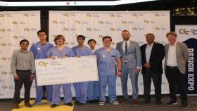
The surgical support device landed Coulter BME its 4th consecutive win for the College of Engineering competition.